
Joint Simulation of Transmission X-ray Imaging on GPU and Patient's Respiration on CPU
Résumé
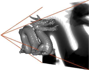
Purpose: We previously proposed to compute the X-ray attenuation from polygons directly on the GPU, using OpenGL, to significantly increase performance without loss of accuracy. The method has been deployed into a training simulator for percutaneous transhepatic cholangiography. The simulations were however restricted to monochromatic X-rays using a point source. They now take into account both the geometrical blur and polychromatic X-rays. Method and Materials: To implement the Beer-Lambert law with a polychromatic beam, additional loops have been included in the simulation pipeline. It is split into rendering passes and uses frame buffer objects to store intermediate results. The source shape is modeled using a variable number of point sources and the incident beam is split into discrete energy channels. The respiration model is composed of ribs, spine, lungs, liver, diaphragm and the external skin. The organ motion simulation is based on anatomical and physiological studies: the model is monitored by two independent active components: the ribs with a kinematics law and the diaphragm tendon with an up and down translation. Other soft-tissue components are passively deformed using a 3D extension of the ChainMail algorithm. The respiration rate is also tunable to modify the respiratory profile. Results: We have extended the simulation pipeline to take into account focal spots that cause geometric unsharpness and polychromatic X-rays, and dynamic polygon meshes of a breathing patient can be used as input data. Conclusions: X-ray transmission images can be fully simulated on the GPU, by using the Beer-Lambert law with polychromatism and taking into account the shape of the source. The respiration of the patient can be modeled to produce dynamic meshes. This is a useful development to improve the level of realism in simulations, when it is needed to retain both speed and accuracy.
Domaines
Modélisation et simulation
Fichier principal
 Vidal2010AAPM-1.pdf (1.33 Mo)
Télécharger le fichier
Vidal2010AAPM-1.pdf (1.33 Mo)
Télécharger le fichier
 no_unsharpness.png (314.52 Ko)
Télécharger le fichier
no_unsharpness.png (314.52 Ko)
Télécharger le fichier
Origine : Fichiers produits par l'(les) auteur(s)
Format : Figure, Image
Loading...