

Quantifying cell polarities in confocal images using 3D wall meshes
Résumé
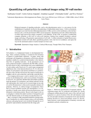
Polarized transport of signaling molecules, such as the phytohormone auxin, is a core process for the establishment of gradients involved in the patterning of multicellular plant tissues. Cell-to-cell polarity of transport can result from the differential deposition of efflux carriers on the membranes at the interface
between cells, as for the pin-formed1 (PIN1) auxin transporter. Quantifying such sub-cellular information in whole organ microscopy images constitutes a real challenge. In this work, we propose a method that computes polarities at the level of cell-to-cell interfaces starting from standard resolution confocal
images and using a 3D geometric representation of cell walls. A robust estimation of spatial fluorescence distribution around cell walls allows quantifying polarity with a fair level of confidence, and opens the way for the automated analysis of polar transport at tissue scale.
Fichier principal
 IAMPS_2019.pdf (1.96 Mo)
Télécharger le fichier
IAMPS_Poster.pdf (16.48 Mo)
Télécharger le fichier
IAMPS_2019.pdf (1.96 Mo)
Télécharger le fichier
IAMPS_Poster.pdf (16.48 Mo)
Télécharger le fichier
Format : Papier court
Origine : Fichiers produits par l'(les) auteur(s)
Loading...